
Researchers have pinpointed the exact number of daily steps needed to dramatically lower the r!sk of a serious health condition
Science Knowledge 24/05/2025 09:24
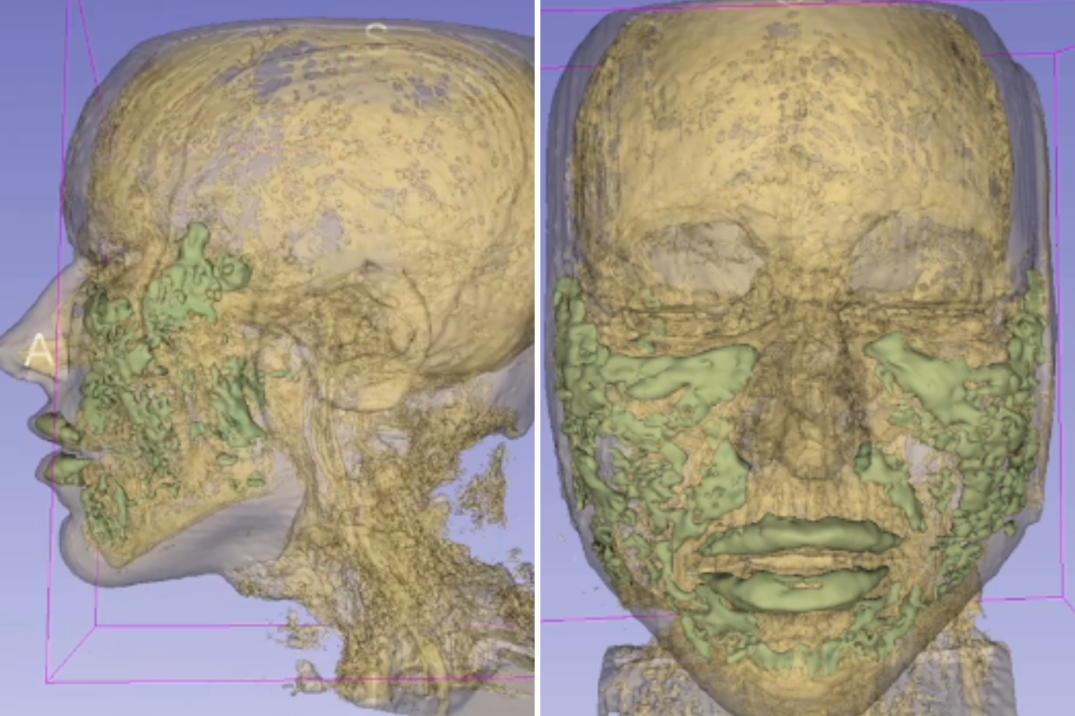
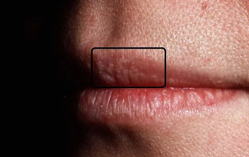
A recent MRI scan of a 33-year-old woman’s face has provided detailed insight into the exact placement of cosmetic fillers after a surgical procedure, highlighting important considerations for aesthetic treatments.
Pictures from the MRI scan. They revealed what filler looks like as it spreads. @kamiparsamd/TikTok
In the realm of cosmetic dermatology, facial fillers have become increasingly popular as a non-surgical method to restore volume, smooth wrinkles, and enhance facial contours. However, understanding exactly where these substances settle post-procedure can be crucial for both practitioners and patients to ensure safety and optimal results.
A recent case involving a 33-year-old woman who underwent facial filler injections was closely examined through an MRI scan, revealing the precise locations of the filler material and shedding light on the behavior of these substances within soft tissues.
The 33-year-old woman underwent a routine cosmetic procedure involving the injection of dermal fillers designed to enhance facial features. Shortly after the procedure, an MRI scan was conducted to assess the filler’s placement and any possible complications.
The magnetic resonance imaging provided a detailed cross-sectional view of the face, clearly showing the distribution of the filler material across various facial layers. This scan allowed clinicians to visualize the exact anatomical positioning of the product, which is typically not visible through conventional examination.
Dr. Jane Roberts, a specialist in dermatologic imaging, explained, "MRI gives us a unique window into the face, enabling us to see exactly where fillers have been placed. This is vital for managing complications or planning further treatments."
While most filler procedures are safe and minimally invasive, complications can occasionally arise if the material migrates or is injected too deeply. Traditional imaging methods often fall short in identifying the filler’s precise location.
MRI offers a non-invasive and radiation-free method to visualize soft tissues with high resolution. This enables healthcare professionals to detect misplaced filler, vascular compromise, or inflammatory responses that might not be apparent otherwise.
The detailed images can guide corrective interventions, such as hyaluronidase injections to dissolve misplaced hyaluronic acid fillers, minimizing risks and improving patient outcomes.
The MRI findings in this case illustrated how fillers integrate within the facial soft tissue. Depending on the type of filler and injection technique, materials may distribute in varied patterns.
Experts note that understanding filler behavior helps in predicting longevity and effects. Some fillers integrate seamlessly, while others may clump or migrate, which can cause uneven results or require corrective measures.
Dr. Roberts noted, "This imaging confirms that fillers do not simply stay in one place; they interact dynamically with surrounding tissue. Knowing this helps clinicians tailor treatments more precisely."
For patients considering cosmetic filler treatments, this case highlights the importance of choosing experienced professionals who understand facial anatomy and utilize safe injection techniques.
Additionally, the availability of MRI imaging as a diagnostic tool provides reassurance and a pathway for addressing any post-procedure concerns.
"It’s essential that patients have access to thorough assessments when complications arise," said Dr. Roberts. "MRI can be an invaluable asset in these situations."
This insightful MRI scan of a 33-year-old woman’s face underscores the critical role of advanced imaging in modern cosmetic dermatology. By revealing the exact placement and behavior of facial fillers, MRI enhances safety, improves treatment planning, and supports better patient care.
As cosmetic procedures continue to evolve, integrating such diagnostic technologies will be key to ensuring both efficacy and safety for patients worldwide.


Every morning, my son handed juice to a garbage man he called “Mr. Tomorrow.” I thought he was a stranger until I learned he held a secret tied to our family.

Discover the chilling secret behind a mother-in-law’s latex gloves in this suspenseful tale of hidden pain, family secrets, and resilience. A gripping story of fear, betrayal, and healing unfolds.

These six powerful juice recipes harness nature’s best ingredients to detoxify, hydrate, and protect your skin—leading to a clearer, firmer, and more luminous complexion.

Singer-rapper reflects on loss and shares heartfelt memories of late “soulmate” Nora Aunor

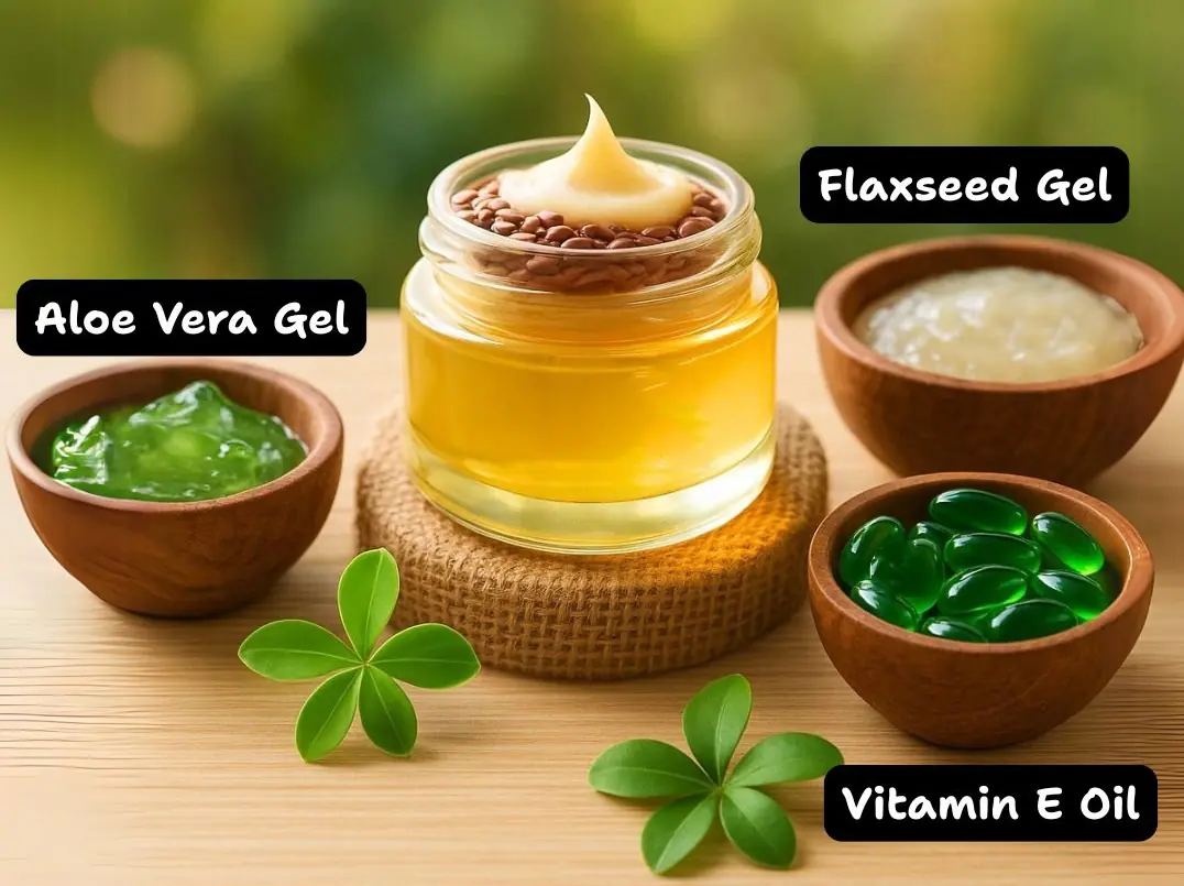
By incorporating this DIY elixir into your daily routine, you nourish your skin deeply, smooth fine lines, and restore a radiant, youthful complexion without exposure to harsh chemicals.

Fennel seed water is more than a refreshing beverage—it’s a holistic health tonic with the power to transform your metabolism, digestive health, skin radiance, and overall vitality.

Actress Lotlot de Leon recently stirred speculation online after posting cryptic messages about lies and truth on her Instagram Stories

Packed with powerful ingredients like cloves, rosemary, garlic, and hibiscus, this oil helps nourish your scalp, stimulate hair follicles, and encourage thicker, healthier hair.

John Rendez expresses deep sorrow over the passing of his dear friend and beloved superstar, Nora Aunor. Despite the passage of time, the loss remains a painful reality that he continues to cope with.

Ruffa Gutierrez opens up about her perspective on marriage and her current relationship with Herbert Bautista

During a playful yet challenging segment on Sarap Di Ba?, Zoren Legaspi faced a tricky question about his ex-girlfriends posed by his wife Carmina Villarroel, leading to a humorous but revealing exchange.

Ready to transform your baking game? These 35 clever hacks will save you time, fix common baking problems, and make your treats taste even better!
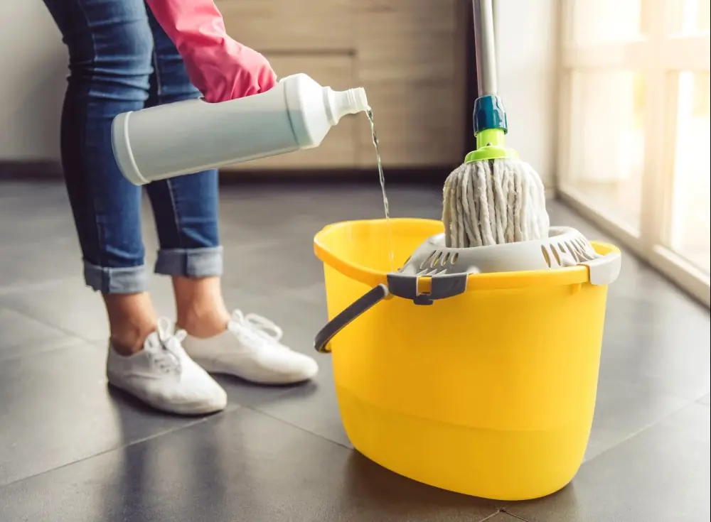
Knowing how frequently to clean your belongings can boost your health and keep your home fresh—discover the best cleaning schedules for everyday items.

A heart-wrenching story of betrayal and love as Polina discovers her husband f@ked his de@th on their wedding day. After the sh0ck and heartbre@k, can she ever trust him again?

Unlock the full potential of your microwave with these clever hacks to save time and make meal prep easier than ever.
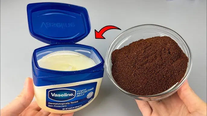
Still, for many, Vaseline offers simple, effective beauty care that doesn’t break the bank. When paired with natural ingredients, it becomes even more powerful - proving that skincare doesn’t have to be complicated to be effective.
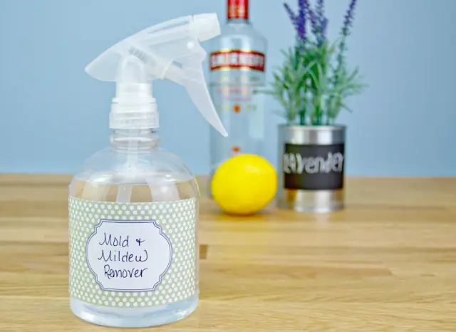
Discover natural, non-toxic ways to eliminate mould from your home using simple ingredients that are just as effective as harsh chemicals.

In a noisy world filled with distractions, embracing silence can restore your focus, clarity, and emotional balance.

After losing her mother, Elanor faces emotional neglect from her husband Jasper, who chooses vacation over support. Her bold response forces change in their fractured marriage. A powerful story of grief, resilience, and reclaiming love.